


 Foundational characteristics of cancer include proliferation, angiogenesis, migration, evasion of apoptosis, and cellular immortality. Find key markers for these cellular processes and antibodies to detect them.
 The SUMOplot™ Analysis Program predicts and scores sumoylation sites in your protein. SUMOylation is a post-translational modification involved in various cellular processes, such as nuclear-cytosolic transport, transcriptional regulation, apoptosis, protein stability, response to stress, and progression through the cell cycle.
 The Autophagy Receptor Motif Plotter predicts and scores autophagy receptor binding sites in your protein. Identifying proteins connected to this pathway is critical to understanding the role of autophagy in physiological as well as pathological processes such as development, differentiation, neurodegenerative diseases, stress, infection, and cancer.
> home > Products > Primary Antibodies > Antibody Collections > GPCR Antibodies > EDG8 Polyclonal Antibody
EDG8 Polyclonal Antibody
Purified Rabbit Polyclonal Antibody (Pab)
- SPECIFICATION
- CITATIONS
- PROTOCOLS
- BACKGROUND

Application
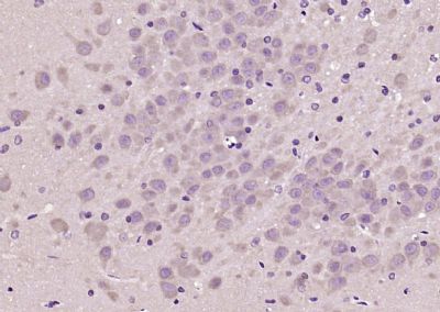
| IHC-P, IHC-F, IF, ICC, E |
|---|---|
| Primary Accession | Q9H228 |
| Reactivity | Rat, Pig, Dog |
| Host | Rabbit |
| Clonality | Polyclonal |
| Calculated MW | 42 KDa |
| Physical State | Liquid |
| Immunogen | KLH conjugated synthetic peptide derived from human EDG8 |
| Epitope Specificity | 1-100/398 |
| Isotype | IgG |
| Purity | affinity purified by Protein A |
| Buffer | 0.01M TBS (pH7.4) with 1% BSA, 0.02% Proclin300 and 50% Glycerol. |
| SUBCELLULAR LOCATION | Cell membrane; Multi-pass membrane protein |
| SIMILARITY | Belongs to the G-protein coupled receptor 1 family. |
| Important Note | This product as supplied is intended for research use only, not for use in human, therapeutic or diagnostic applications. |
| Background Descriptions | The EDG (endothelial differentiation gene) family of G protein coupled receptors consists of eight family members that bind lysophospholipid (LPL) mediators, including sphingosine-1-phosphate (SPP) and lysophosphatidic acid (LPA). EDG-1, EDG-3, EDG-5 (also designated H218 and AGR16) and EDG-8 bind SPP with high affinity. EDG-6 is a low affinity receptor for SPP. LPA preferentially binds to EDG-2, EDG-4 and EDG-7. The EDG receptors couple to multiple G proteins to signal through Ras, MAP kinase, Rho, Phospholipase C or other tyrosine kinases, which lead to cell survival, growth, migration and differentiation. EDG-1 signals through Gi proteins to activate Akt and is expressed in glioma cells. EDG-2 is expressed in brain, especially in white matter tract regions, while EDG-3 is expressed in cardiovascular tissue and in cerebellum. EDG-4 is highly expressed on leukocytes and brain, and EDG-5 has wide tissue distribution, including cardiovascular tissue and brain. EDG-6, which is expressed in lymphoid and hematopoietic tissues and in lung, signals through Gi/o proteins, which activate growth related pathways. |
| Gene ID | 53637 |
|---|---|
| Other Names | Sphingosine 1-phosphate receptor 5, S1P receptor 5, S1P5, Endothelial differentiation G-protein-coupled receptor 8, Sphingosine 1-phosphate receptor Edg-8, S1P receptor Edg-8, S1PR5, EDG8 |
| Target/Specificity | Widely expressed in the brain, most prominently in the corpus callosum, which is predominantly white matter. Detected in spleen, peripheral blood leukocytes, placenta, lung, aorta and fetal spleen. Low-level signal detected in many tissue extracts. Overexpressed in leukemic large granular lymphocytes. Isoform 1 is predominantly expressed in peripheral tissues. Isoform 2 is expressed in brain, spleen and peripheral blood leukocytes. |
| Dilution | IHC-P=1:100-500,IHC-F=1:100-500,ICC=1:100-500,IF=1:100-500,ELISA=1:5000-10000 |
| Format | 0.01M TBS(pH7.4), 0.09% (W/V) sodium azide and 50% Glyce |
| Storage | Store at -20 ℃ for one year. Avoid repeated freeze/thaw cycles. When reconstituted in sterile pH 7.4 0.01M PBS or diluent of antibody the antibody is stable for at least two weeks at 2-4 ℃. |
| Name | S1PR5 |
|---|---|
| Synonyms | EDG8 |
| Function | Receptor for the lysosphingolipid sphingosine 1-phosphate (S1P). S1P is a bioactive lysophospholipid that elicits diverse physiological effect on most types of cells and tissues. Is coupled to both the G(i/0)alpha and G(12) subclass of heteromeric G-proteins (By similarity). May play a regulatory role in the transformation of radial glial cells into astrocytes and may affect proliferative activity of these cells. |
| Cellular Location | Cell membrane; Multi-pass membrane protein. |
| Tissue Location | Widely expressed in the brain, most prominently in the corpus callosum, which is predominantly white matter. Detected in spleen, peripheral blood leukocytes, placenta, lung, aorta and fetal spleen. Low-level signal detected in many tissue extracts Overexpressed in leukemic large granular lymphocytes. Isoform 1 is predominantly expressed in peripheral tissues. Isoform 2 is expressed in brain, spleen and peripheral blood leukocytes |
Research Areas
Citations (0)

Thousands of laboratories across the world have published research that depended on the performance of antibodies from Abcepta to advance their research. Check out links to articles that cite our products in major peer-reviewed journals, organized by research category.
Submit your citation using an Abcepta antibody to
info@abcepta.com, and receive a free "I Love Antibodies" mug.
info@abcepta.com, and receive a free "I Love Antibodies" mug.

Application Protocols
Provided below are standard protocols that you may find useful for product applications.
Abcepta welcomes feedback from its customers.
If you have used an Abcepta product and would like to share how it has performed, please click on the "Submit Review" button and provide the requested information. Our staff will examine and post your review and contact you if needed.
If you have any additional inquiries please email technical services at tech@abcepta.com.
$ 385.00
Cat# AP57973
Ordering Information
United States
AlbaniaAustraliaAustriaBelgiumBosnia & HerzegovinaBrazilBulgariaCanadaCentral AmericaChinaCroatiaCyprusCzech RepublicDenmarkEstoniaFinlandFranceGermanyGreeceHong KongHungaryIcelandIndiaIndonesiaIrelandIsraelItalyJapanLatviaLithuaniaLuxembourgMacedoniaMalaysiaMaltaMexicoNetherlandsNew ZealandNorwayPakistanPolandPortugalRomaniaSerbiaSingaporeSlovakiaSloveniaSouth AfricaSouth KoreaSpainSwedenSwitzerlandTaiwanTurkeyUnited KingdomUnited StatesVietnamWorldwideOthers
USA Headquarters
(888) 735-7227 / (858) 622-0099 or (858) 875-1900
Other Products
Shipping Information
Domestic orders (in stock items)
Shipped out the same day. Orders placed after 1 PM (PST) will ship out the next business day.
International orders
Contact your local distributors
